|
SEMG: The Spinal Scan
Surface Electromyographic Analysis
42 Point Neurological Analysis of Your Spine
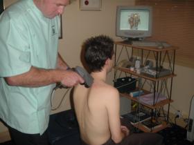
During the initial consultation patients usually have a detailed Postural Evaluation and Spinal Scan – Surface Electromyographic Study (SEMG).
The SEMG precisely measures the nerve signals passing out of your spine to the important postural muscles of your back. The measurement is very precise – to one tenth of a microvolt.
Essentially, we are measuring the electrical potential of the muscles within your spine. The MOTOR function of the nervous system.
This state of the art technology will objectively help us:
- Detect areas of nerve disturbance
- Detect areas of muscle imbalance
- Detect areas of postural disturbance or imbalance
- Detect spinal problems in children and adolescents
- Document and monitor your results
- Help us to deliver the appropriate Chiropractic Care
SEMG – Surface Electromyography
Nerves control the muscles of the spine. The SEMG measures how well the motor nerves are working by reading the amount of current found in the muscles.
Structural damage, misalignments, scoliosis, disc injuries or postural imbalances disturb the function of the nerves. This can cause an abnormal amount of muscle activity, muscle tightness or even muscle spasm, fibrillation and spasticity.
Different colours show the amount of muscle activity on the scan. It is very easy to understand and correlate it to problem areas within the spine, or areas that still need more work.
The results are shown as coloured bars on the print out:
- White; show normal nerve and muscle function, within normal limits
- Green; mild nerve interference and muscle tightness
- Blue; moderate nerve interference and muscle spasm
- Red; severe nerve irritation and muscle spasm; 3 standard deviations above normal
- Black; nerve damage and irritation with extreme muscle spasm
- Yellow; nerve compression (shut down), neurogenic muscle weakness or wasting
Muscle Balance or Symmetry
Muscle balance is extremely important because the vertebrae in the spine depend on the muscles to move properly. If one or more vertebra do not move properly this will disturb nerve function. Nerve interference or disturbance creates muscle imbalances.
Any abnormal muscle pattern or “asymmetry pattern” reveals an increased amount of tension or pull on one side compared to the other. This “tug of war” within the muscles must be removed to ensure good spinal function. Relapses are inevitable if asymmetry patterns are left within the spine.
The question of “How do I know I am getting the right amount care?” is answered with the use of this scanning technology. You will be able to see the changes clearly on progressive scans. With this technology we will be able to provide a “tailor-made” program to meet your specific health needs.
If you had a Computer Spinal Scan then the results of your analysis will be given to you on your next appointment – The Report of Clinical Findings or Dr’s Report.
SEMG Result Prior To Care
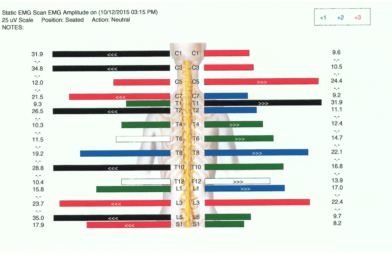 The first scan on the right is an example of a patient in very bad shape at the beginning of care.
The large red and black bars show multiple areas of nerve interference and potential nerve damage.
Note: The SEMG does not measure pain but nerve damage and altered muscle function (muscle spasm).
The more reds or blacks in an area of your spine => The more likely you are to have pain and disability in that area.
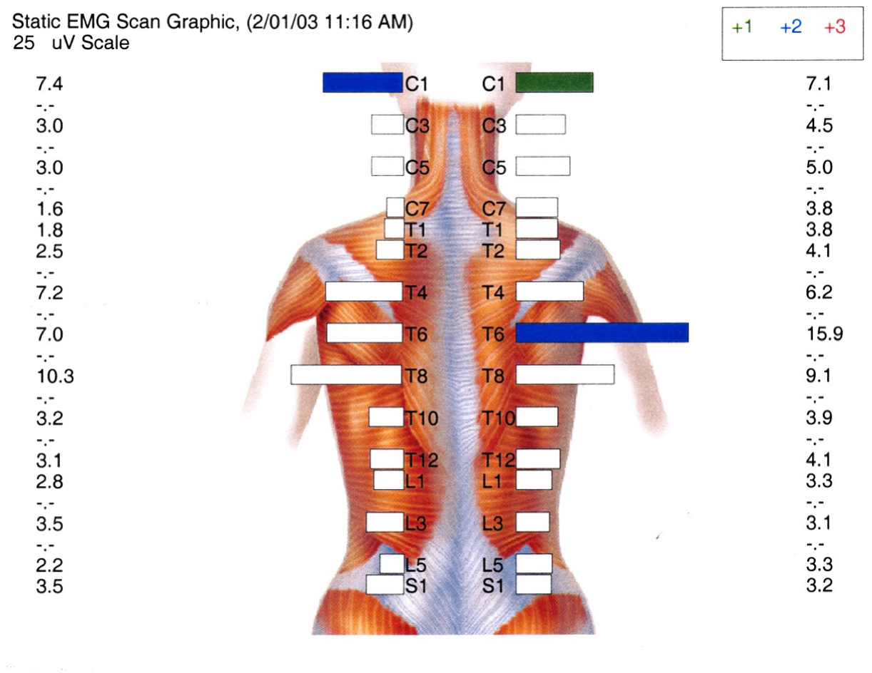 SEMG After Spinal Reconstructive Care
The second scan is an example of a patient who has successfully completed their treatment protocol and really participated in their recovery with corrective exercises and appropriate stretching.
The spine is stable and quite symmetrical.
The white bars show the electrical potential of the muscle within an optimal range.
Good spinal function is essential for better health.